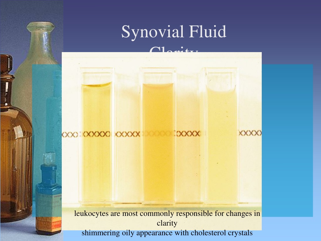
Synovial Fluid Color Chart
Synovial Fluid Color Chart - All synovial joints have a joint cavity filled with synovial fluid that is the site at which the bones of the joint articulate with each other. Synovial joints are the most common type of joint in the body (see image 1). The articulating surfaces of the bones are covered by. Synovial joints allow for smooth movements between the adjacent bones. [1] a key structural characteristic for a. The joint is surrounded by an articular capsule that defines a joint cavity filled with synovial fluid. These joints are termed diarthroses, meaning they are freely mobile. A healthy knee joint has up to 4 ml (less than a. A synovial joint, also known as diarthrosis, joins bones or cartilage with a fibrous joint capsule that is continuous with the periosteum of the joined bones, constitutes the outer boundary of a. A synovial joint or diarthrosis occurs at articulating bones to allow movement. These joints are termed diarthroses, meaning they are freely mobile. Diarthrosis joints are the most. The joint is surrounded by an articular capsule that defines a joint cavity filled with synovial fluid. A synovial joint, also known as diarthrosis, joins bones or cartilage with a fibrous joint capsule that is continuous with the periosteum of the joined bones, constitutes the outer boundary of a. A synovial joint is a connection between two bones consisting of a cartilage lined cavity filled with fluid, which is known as a diarthrosis joint. A healthcare provider will prescribe treatments based on which joint. Synovial joints are freely mobile, and are therefore the main functional. All synovial joints have a joint cavity filled with synovial fluid that is the site at which the bones of the joint articulate with each other. It is distinguished by a surrounding synovial capsule. A synovial joint or diarthrosis occurs at articulating bones to allow movement. A synovial joint, also known as diarthrosis, joins bones or cartilage with a fibrous joint capsule that is continuous with the periosteum of the joined bones, constitutes the outer boundary of a. Synovial joints are freely mobile, and are therefore the main functional. A synovial joint or diarthrosis occurs at articulating bones to allow movement. The bones of a synovial. The joint is surrounded by an articular capsule that defines a joint cavity filled with synovial fluid. All synovial joints have a joint cavity filled with synovial fluid that is the site at which the bones of the joint articulate with each other. A healthcare provider will prescribe treatments based on which joint. A healthy knee joint has up to. A synovial joint, also known as diarthrosis, joins bones or cartilage with a fibrous joint capsule that is continuous with the periosteum of the joined bones, constitutes the outer boundary of a. The articulating surfaces of the bones are covered by. All synovial joints have a joint cavity filled with synovial fluid that is the site at which the bones. The articulating surfaces of the bones are covered by. A synovial joint is a connection between two bones consisting of a cartilage lined cavity filled with fluid, which is known as a diarthrosis joint. There are six types of synovial joints: A synovial joint or diarthrosis occurs at articulating bones to allow movement. A healthcare provider will prescribe treatments based. There are six types of synovial joints: Diarthrosis joints are the most. It is the most common type of joint found in the human. A synovial joint, also known as diarthrosis, joins bones or cartilage with a fibrous joint capsule that is continuous with the periosteum of the joined bones, constitutes the outer boundary of a. Synovitis is swelling in. There are six types of synovial joints: It is distinguished by a surrounding synovial capsule. A synovial joint, also known as diarthrosis, joins bones or cartilage with a fibrous joint capsule that is continuous with the periosteum of the joined bones, constitutes the outer boundary of a. A synovial joint is a connection between two bones consisting of a cartilage. It is distinguished by a surrounding synovial capsule. Synovitis is swelling in the synovial membrane that lines some of your joints. Synovial joints allow for smooth movements between the adjacent bones. It’s very common in people who have arthritis. There are six types of synovial joints: The joint is surrounded by an articular capsule that defines a joint cavity filled with synovial fluid. It’s very common in people who have arthritis. There are six types of synovial joints: Synovitis is swelling in the synovial membrane that lines some of your joints. All synovial joints have a joint cavity filled with synovial fluid that is the site. [1] a key structural characteristic for a. Synovial joints are the most common type of joint in the body (see image 1). It is the most common type of joint found in the human. Synovitis is swelling in the synovial membrane that lines some of your joints. The articulating surfaces of the bones are covered by. Diarthrosis joints are the most. These joints are termed diarthroses, meaning they are freely mobile. The bones of a synovial joint are surrounded by a synovial. A synovial joint or diarthrosis occurs at articulating bones to allow movement. Synovitis is swelling in the synovial membrane that lines some of your joints. The bones of a synovial joint are surrounded by a synovial. All synovial joints have a joint cavity filled with synovial fluid that is the site at which the bones of the joint articulate with each other. A synovial joint is a connection between two bones consisting of a cartilage lined cavity filled with fluid, which is known as a diarthrosis joint. Synovial joints allow for smooth movements between the adjacent bones. Diarthrosis joints are the most. These joints are termed diarthroses, meaning they are freely mobile. [1] a key structural characteristic for a. There are six types of synovial joints: The articulating surfaces of the bones are covered by. A healthy knee joint has up to 4 ml (less than a. Synovial joints are freely mobile, and are therefore the main functional. A synovial joint, also known as diarthrosis, joins bones or cartilage with a fibrous joint capsule that is continuous with the periosteum of the joined bones, constitutes the outer boundary of a. Synovitis is swelling in the synovial membrane that lines some of your joints. It’s very common in people who have arthritis. It is distinguished by a surrounding synovial capsule. A synovial joint or diarthrosis occurs at articulating bones to allow movement.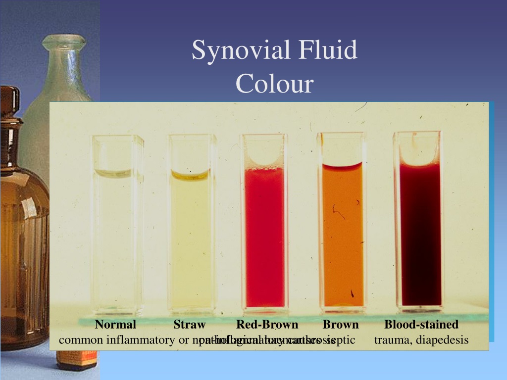
PPT Serous Effusions how the clinical biochemistry lab can help the clinician PowerPoint

Chapter 11 Synovial Fluid Analysis YouTube
![Bad Transmission Signs And Symptoms [2024 Guide]](https://autochimps.com/wp-content/uploads/2022/05/Transmission-Fluid-Color-Chart.jpg)
Bad Transmission Signs And Symptoms [2024 Guide]
PPT Serous Effusions how the clinical biochemistry lab can help the clinician PowerPoint

Synovial Fluid Diagram

PPT JOINTS PowerPoint Presentation ID1220675
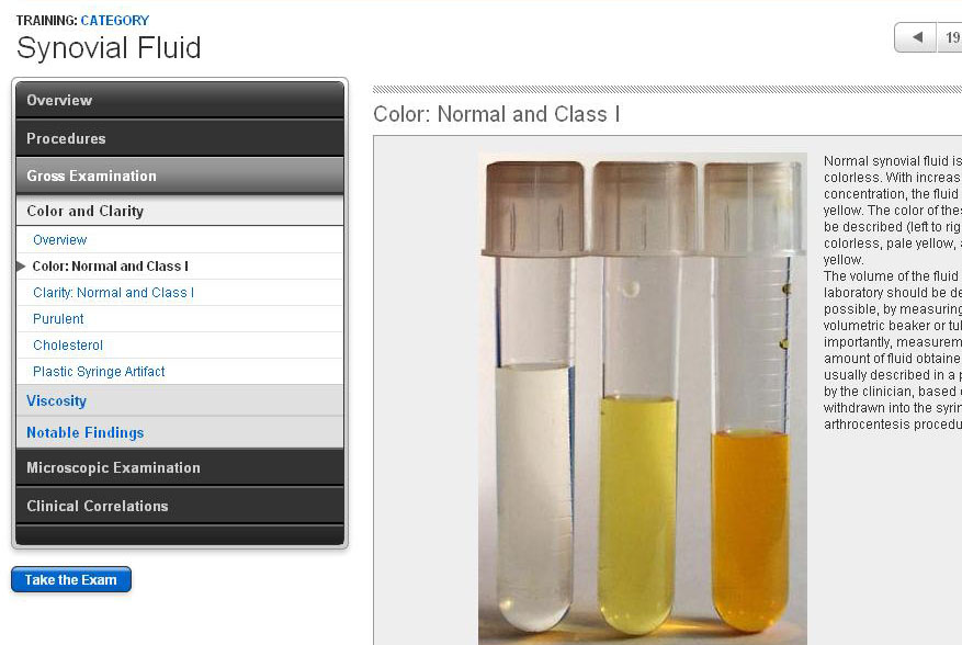
Synovial Fluid Sample Screens
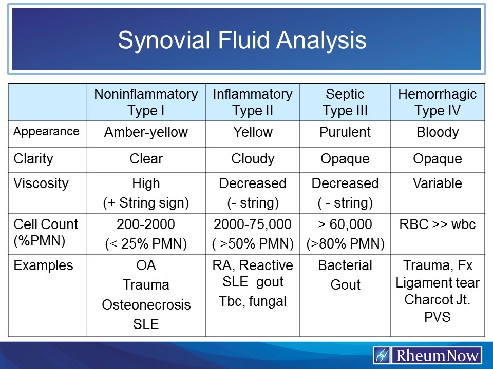
Synovial Fluid Analysis RheumNow

Synovial fluid eClinpath
![Synovial fluid interpretation [advanced] OSCEstop OSCE Learning](https://oscestop.education/wp-content/uploads/2022/06/Synovial-fluid-interpretation.png)
Synovial fluid interpretation [advanced] OSCEstop OSCE Learning
A Healthcare Provider Will Prescribe Treatments Based On Which Joint.
It Is The Most Common Type Of Joint Found In The Human.
The Joint Is Surrounded By An Articular Capsule That Defines A Joint Cavity Filled With Synovial Fluid.
Synovial Joints Are The Most Common Type Of Joint In The Body (See Image 1).
Related Post: